Blue Monomacra Flea Beetle (Monomacra violacea)
The blue flea beetle may be found eating holes in the leaves of most Passiflora species, often in groups of 5-15. Host species include some highly cyanogenic species such as costaricensis and pittieri, indicating that M. violacea, like its congener M. chontalensis, may be able to tolerate cyanide-releasing chemicals in its diet. Its wide distribution at La Selva is somewhat like Heliconius cydno, the most "generalist" of the La Selva heliconiines. Also like H. cydno, this species is frequently found in forest light gaps and clearings as well as forest edge habitats. Unlike H. cydno, M. violacea seems highly successful on P. lobata, the Passiflora with hooked trichomes. In fact, most P. lobata "infested" with a population of M. violacea die within 6 month to a year.. This species may be partially responsible for the high demographic turnover rate for P. lobata.
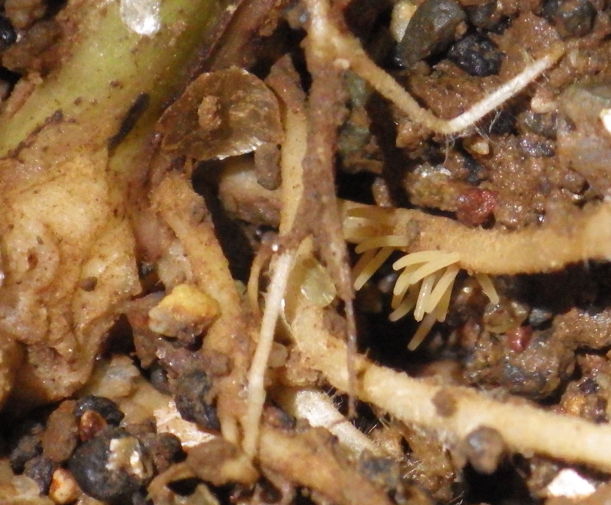
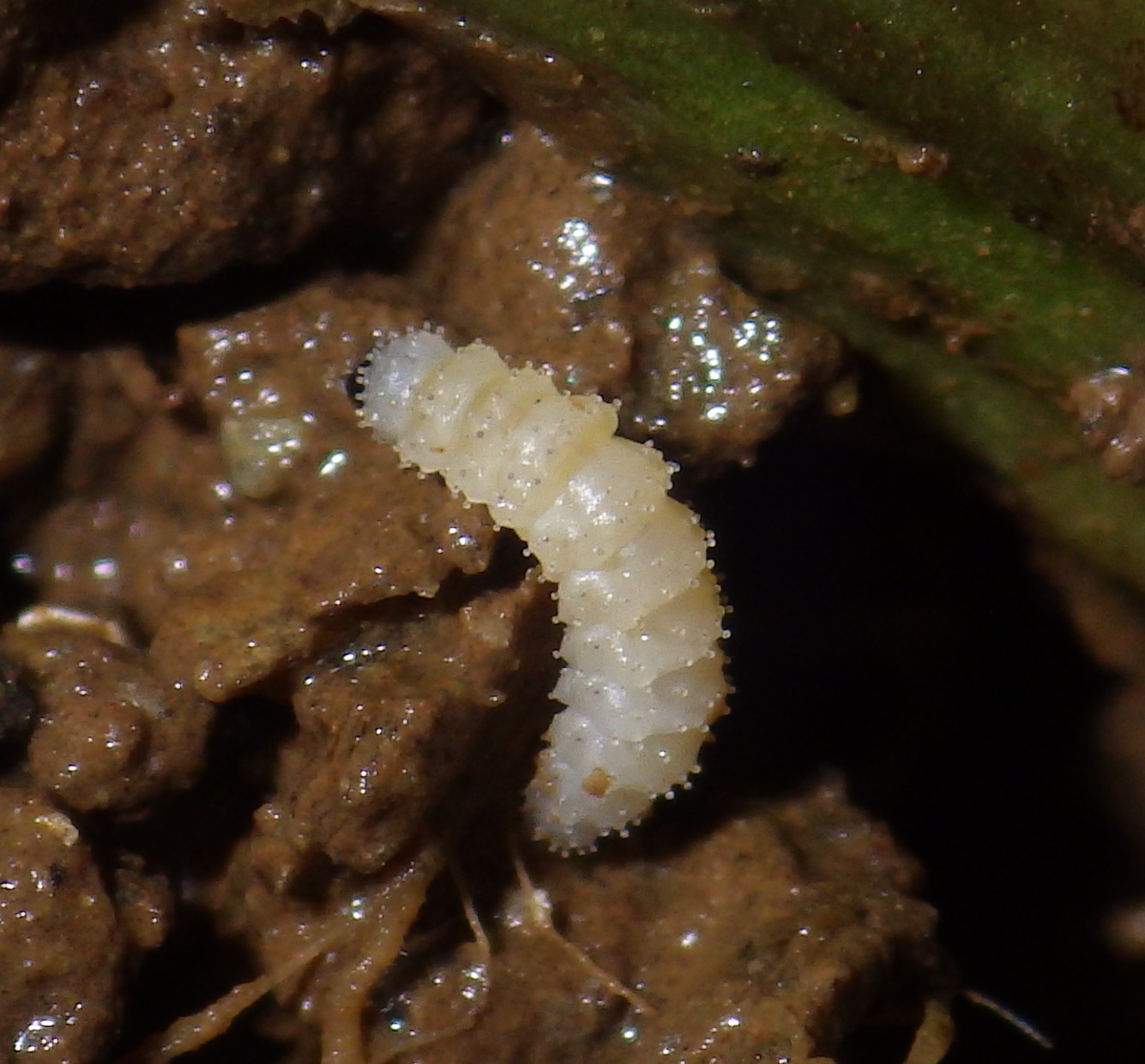
Eggs and larvae of this species resemble those of the closely related Parchicola, although the eggs are attached at end of the cylinder rather than at the side as in Parchicola. Like Parchicola, M. violacea larvae possess setae on the dorsal and lateral lobes, modified into spherical bodies ("balloon organs") which look as if they might store and/or dispense a chemical defense. They also appear to be superficial root feeders and have not yet been been found eating stem tissues. Caged populations in the shadehouse isolated with P. vitifolia and P. auriculata did not reproduce after 2-3 months, and only P. lobata has proven to be a successful larval host to date. This taken with the fact that after the very wet season of May-August 2015 I could only find this species on P. lobata, suggests that lobata may be the primary and/or preferred larval host. The numerous sightings on other Passiflora species perhaps indicate adult host preference only. In this respect it is interesting to note that, among Passiflora species, P. lobata may possess the smallest amount of cyanogenic glycosides (see Figure 4 and Figure 6).
Common in all seasons of the year, often in groups of 10-20 on a single host plant. Mating takes place on the leaves of the host plant. Eggs are laid on rootlets at ground level or 1cm into loose soil around the base of the host plant. Eggs hatch into unpigmented nearly transparent larvae covered with tiny paddle-shaped flaps, with two rows of flaps on each segment. This contrasts with similar larvae of Parchicola, which have only one row per segment. Larvae feed on rootlets by chewing pits into the pith, avoiding the hard core of the root. They grow slowly in captivity and are still in the first instar after 6 days. The larvae seem to retain the pigmented "flap" organs throughout this instar. However, newly moulted second instar larvae possess unpigmented spherical balloon organs (see photos below) that are presumably derived from the flap organs during the moult. A third instar larva of unknown age (photos below), but not newly moulted, was still feeding after 6 days. It then began wandering for 8 days, presumably seeking a pupation site. Assuming the second instar also lasts at least 6 days, in would appear that the larval stage takes at least 18 days to complete development, perhaps much longer.
Like genus Parchicola, Monomacra has a flattened, incised elongated rectangle on the hind edge of the pronotum. It is difficult to see in these photos because of the reflectivity of the cuticle. This species is readily recognizable in museum collections by the shiny blue cuticle.